Neuroscience
Our Brain’s Conductor
The cerebellum has been underestimated for a long time. However, the results of two research groups in the Collaborative Research Center show that it plays a crucial role in regulating emotions.
Brushing your teeth, riding a bike, eating an apple: We are only able to perform these everyday actions thanks to an often-overlooked region of the brain known as the cerebellum. It weighs just 150 grams, but contains 80 per cent of our neurons. As a motor control center, it controls key motion sequences in the body and is responsible for our balance and coordination. It has been known for several years that the cerebellum also controls cognitive processes such as problem solving. And not only that. For a long time, the fact that the cerebellum also plays an important role in regulating our emotions – such as when processing fear – has been ignored. Professor Melanie Mark from Ruhr University Bochum and Professor Dagmar Timmann from the University of Duisburg-Essen are two of the first researchers to provide experimental evidence that the cerebellum contributes towards both the learning and the extinction of conditioned fear responses.
To understand fear learning
To investigate the role that the cerebellum plays in fear learning, the two neuroscientists conducted learning experiments – the neurologist with humans and the neurobiologist with mice. “In our studies, we draw on classic fear-conditioning experiments and compare healthy humans and mice with those that have a cerebellar disease, ataxia,” says Timmann, summarizing the shared study design.
Ataxia
In her latest study, the clinical neuroscientist selected 20 subjects who suffer from rare cerebellar diseases such as spinocerebellar ataxia type 6 (SCA6), in addition to 20 healthy people. “The movement disorder SCA6 is triggered by a genetic defect similar to Huntington’s disease and only affects a very small number of people in Germany,” explains Timmann, who has been offering ataxia consultations at Essen University Hospital for many years. “SCA6 is associated with a loss of a specific type of neuron in the cerebellum, the Purkinje cells. The Purkinje cells are important as intermediaries between the cerebellum and the rest of the brain. For instance, the cerebellum helps the cerebrum to optimize motion sequences,” says the researcher.
Fear-conditioning experiments
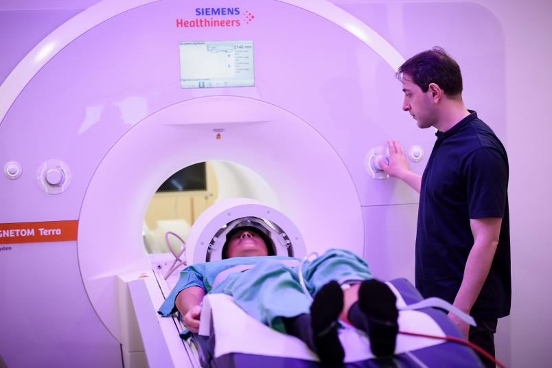
In their study, Timmann and her team had patients and participants learn and then unlearn fear within two days while observing them in the 7 tesla MRI scanner.
7 tesla MRI system
To trigger fear, the subjects were given an unpleasant electric shock on their shin whenever they saw a certain image on the monitor on day 1. They soon learned that an electric shock was imminent whenever this image was shown. The skin conductance and pupil size of the subjects were measured during the experiment. “Our subjects’ pupils dilated and their skin conductance increased when the shock took place. And, as shown over the course of the experiment, also when pain was expected,” says Timmann. The observation corresponds to the answers that the subjects gave in the questionnaires at the end of the experiment. While the image triggered neutral feelings at the start of the experiment, the people stated at the end that the image triggered fear in them. The image was shown again on day 2, but this time it was not followed by a shock. “The subjects unlearned the fear response, which is called extinction,” explains Timmann.
Deficits when learning and unlearning fear
And the result? The direct comparison of healthy subjects and people with ataxia confirmed the assumption that people with ataxia have deficits when learning and unlearning fear. Not only did the acquisition and consolidation of the learned fear response take longer than in the healthy control group, but unlearning the fear was also more prolonged. However, the deficits were much lower than expected: “Beforehand, we assumed that our ataxia patients would be much more significantly impaired during the fear conditioning and that this, in turn, would be associated with clearly visible changes in the cerebellum,” says Timmann. However, in the 7 tesla MRI, the activation pattern in the ataxia patients was also reasonably well preserved and only showed minor deviations from the healthy participants.
Mouse models
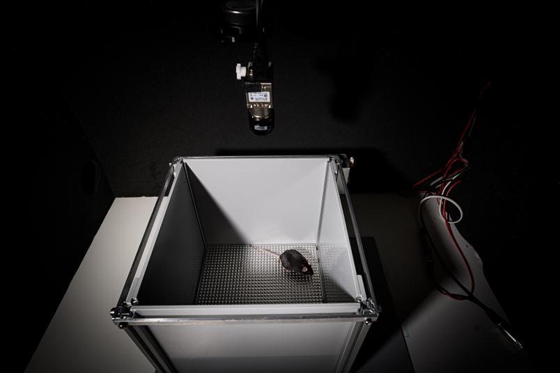
To confirm the Essen clinician’s observations, her research colleague in Bochum, neurobiologist Melanie Mark, conducted the fear conditioning study with healthy mice and mice suffering from SCA6. Mark used SCA6 mouse models that she had already developed for previous studies.
The mouse model
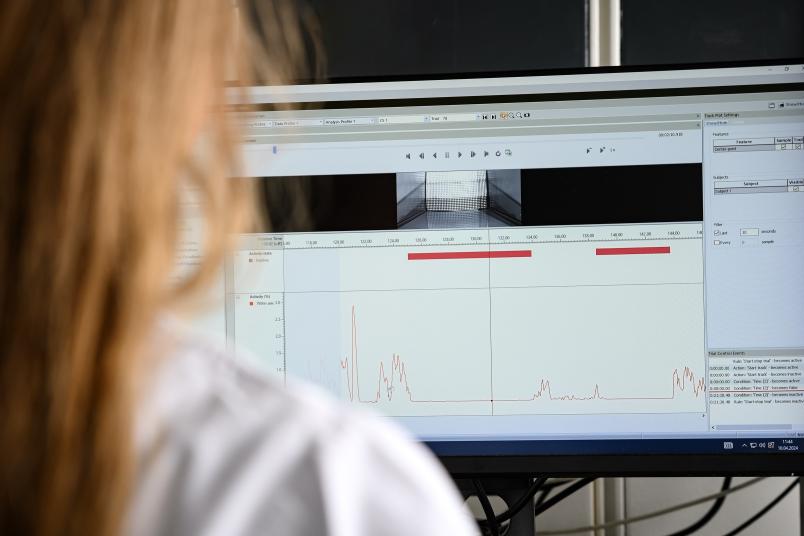
“We wanted to find out whether our degenerative cerebellum mouse model shows similar deficits in fear conditioning,” says Mark. During the learning experiment, the healthy and sick mice were conditioned on day 1 by giving them a shock whenever they heard a certain tone, causing them to learn to associate the tone with the unpleasant shock from then on. Whenever they heard the tone after that, both the healthy and the sick mice froze in fear of the electric shock.

Our SCA6 mice did not consolidate what they had learned.
One day later, only the tone sounded, but there was no shock. The healthy mice nevertheless froze in fear as they expected the shock. In contrast to this, the SCA6 mice showed much less fear on day 2. “Our SCA6 mice were able to learn the fear response exactly like the people with ataxia, but they did not consolidate what they had learned. Their memory of the learned association task did not last until the next day,” explains Mark. The researcher was thus able to show that the fear memory in the SCA6 mice was disrupted in comparison to the healthy mice. The cerebellar disease prevented the mice from consolidating what they had learned and, based on this, from being able to make a learned prediction.

This is desirable from an evolutionary perspective.
Mark thus came to the same conclusion as Timmann: The cerebellum plays a role in learning fear responses. However, the deficits were also much lower than expected in the mouse model. “In this chronic illness, other regions of the brain have possibly learned to compensate for the cerebellar deficit. This is desirable from an evolutionary perspective. If a region fails, the whole neuronal circuit does not immediately collapse. This does not mean that the cerebellum is not involved,” explain Mark and Timmann.

We can specifically control and stimulate individual cells and cell populations in the cerebellum.
Melanie Mark’s team is now working hard on rectifying the learning deficits in SCA 6 mice using various methods. “The special thing about our mouse model is that we can specifically control and stimulate individual cells and cell populations in the cerebellum to see what role they play in learning and forgetting fear,” explains Mark. In the long term, Mark and Timmann hope to better understand the precise contribution made by the cerebellum in these learning processes. The special cooperation between neurology and neurobiology, which the Collaborative Research Center 1280 makes possible, is indispensable here.
Symphonies of thoughts and emotions
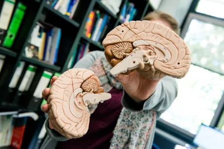
With their research, Mark and Timmann are paying a region of the brain the attention it has long deserved. Their results confirm that the cerebellum plays an important role in fine-tuning our fear responses. “The cerebellum is our brain’s conductor, which controls the symphonies of our thoughts and emotions,” describes Mark. “It collects and organizes all of the information and then passes on the knowledge to other regions of the brain and makes a prediction.”

