Neurobiology
Switching Off Fear
The extended amygdala plays a major role in assessing diffuse threats and in persistent fear reactions. Studies show that it is also involved in learning and unlearning specific fear stimuli.
If a neighbor’s dog dislikes children and jumps up at them, barking loudly, the children might give all dogs a wide berth for a while. A sensible learning effect – as who knows whether all dogs are so aggressive? You can also free yourself of a fear learned in this way: If the children have better experiences with friendlier dogs, they can lose their fear again. “For some conditions, however, this unlearning of the fear response doesn’t work correctly,” says Dr. Katharina Spoida from the Department of General Zoology and Neurobiology at Ruhr University Bochum. Following a traumatic experience, those affected are unable to break the link between a neutral stimulus and fear. They become overwhelmed by overpowering anxiety in everyday life – signs of post-traumatic stress disorder.
Katharina Spoida wants to know what happens in the brain when a fear memory is formed and how this fear is later extinguished. The focus lies on an area of the brain that we know plays an important role in the development and rejection of fear: the amygdala. The neurotransmitter serotonin and, with it, the receptors via which it can transmit signals to neurons also play a key role.

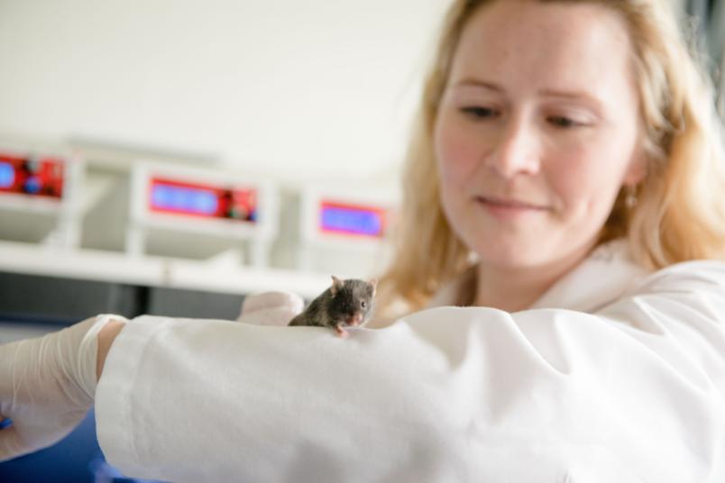
The mice used by the researchers are derived from genetically modified mouse lines.
To find out which processes take place in the brain during the learning and unlearning of fear, the researchers are using various mouse models. “There are genetically modified mice that lack a certain serotonin receptor called 5-HT2C,” reports Katharina Spoida. These knock-out mice are known to differ from wild mice in that, among other things, they are less anxious. Spoida’s team is investigating how fear is learned and unlearned in the mice in comparative experiments.
They presented the various mice with a 30-second tone as a neutral stimulus and followed this with an unpleasant but painless electrical stimulus. “It only takes five repetitions for the mice to learn this link,” reports the researcher. “We can tell this because the mice display a behavior that we call freezing, a motionless pause, after the tone is played. In a threatening situation, freezing represents an important survival strategy, as it makes the mice less visible to the predator. On the following day, the researchers played the corresponding tone to the animals several times without it being followed by the unpleasant stimulus. “The remarkable thing was that the knock-out mice learned to no longer fear the tone much faster than mice without the genetic modification,” says Katharina Spoida. The absence of the serotonin receptor, therefore, appears to be an advantage for extinction learning.
The researchers investigated this phenomenon further and found that the knock-out mice display changes in their neuronal activity in two different areas of the brain. This includes a specific sub-region of the dorsal raphe nucleus (DRN), which is generally the main location for serotonin production in our brain. The researchers also discovered deviating neuronal activity in the bed nucleus of the stria terminalis (BNST), which is part of the extended amygdala. The research results also show a connection between both regions of the brain, suggesting that an interaction could be important for improved extinction learning.
Knock-out mice learned more quickly
“The interesting thing about this experiment is that the results look completely different when carried out with females,” reports Katharina Spoida. “We do not see the altered learning affects in them.” This finding is of particular importance, as twice as many women as men suffer from post-traumatic stress disorder. However, up to now, everyone receives the same treatment.
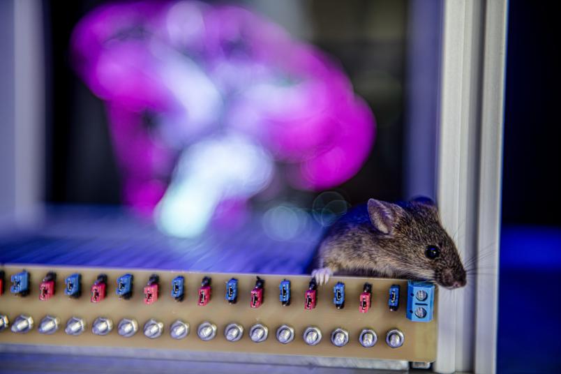
To uncover the reasons for the change in learning in male knock-out mice and gain general insights into signal processing during the learning of fear, the researchers at the Department of General Zoology and Neurobiology are using targeted measures to trigger neuron activity themselves. This can be achieved using light or chemical stimuli.
Optogenetics
“This approach enables us to activate or inhibit cells in a targeted manner and see what effect this has on the animal’s learning of fear,” explains Katharina Spoida. If they inhibit a subset of corticotropin-releasing factor (CRF) neurons in knock-out mice in the BNST, these mice unlearn the fear more slowly. If they activate them in wildtype mice, the unlearning effect is accelerated. The research team was thus able to confirm which regions of the brain contain the crucial structures for learning and unlearning fear in their mouse model. “The bed nucleus of the stria terminalis is split into areas that tend to promote fear and those that tend to reduce fear,” says the researcher. In male knock-out mice, activity in the fear-reducing area is increased compared to their wild counterparts, and lower in the fear-promoting areas. The absence of the 5-HT2C receptor appears to push neuronal activity in the BNST in an extinction-supporting direction, and the CRF neurons play an important role in this.

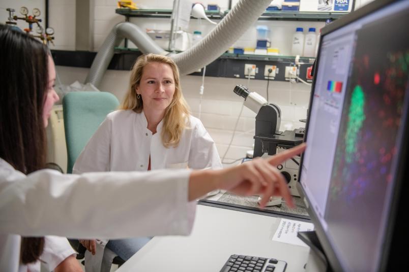
Katharina Spoida, Sandra Süß and their team can directly activate and inhibit cells. This enables extremely detailed research into learning phenomena at cellular level.
In view of the medicinal treatment of patients with post-traumatic stress disorder, the findings from fundamental research explain why symptoms often tend to worsen rather than improve in the beginning: By using what are known as selective serotonin reuptake inhibitors (SSRIs), more serotonin is freely available in the brain and can activate the various fear-promoting serotonin receptors. Only after several weeks do the symptoms improve, as the cells withdraw the receptors due to the constant overstimulation. “These initial effects could be minimized by combining medications to block the receptors,” says Katharina Spoida. In general, she hopes to help make the treatment more specific in the future. “In the future, we should take greater account of gender-specific differences when researching and treating mental illnesses,” she says with certainty.
