
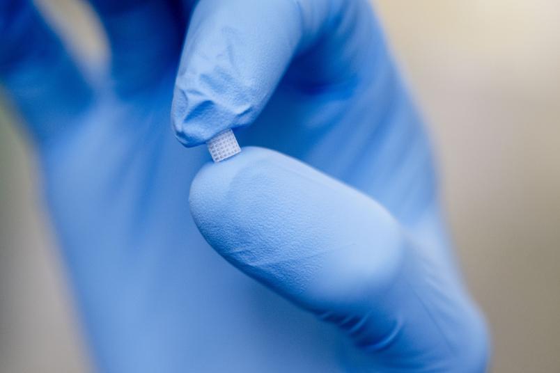
The researchers derived the structure of human bones from computer tomography imaging and replicated it with a 3D printer.
3R Principle
Studying Bone Metabolism on a Chip
Why do artificial joints come loose at some point? A research team is seeking to answer this question. Highly developed cell culture systems and bioreactors are being used for this instead of animal experimentation.
Over 400,000 individuals in Germany receive an artificial knee or hip joint every year. This is one of the most common orthopedic operations and usually alleviates patients’ issues quickly, although an implant often has to be replaced after 12 to 15 years on average. In Germany, similar to other European countries, about 21 percent of artificial joints require adjustment each year, based on registry data.
“One reason for this is that the prosthetic begins to loosen at some point,” explains Dr. Jochen Salber, Director of Experimental Surgery at Knappschaft Kliniken Universitätsklinikum Bochum. This loosening stems from complex processes at the interface between the metal implant and the surrounding bone tissue. The role played by metallic nanoparticles that break away from the implant when the artificial joint moves, as well as the surface consistency of the implant itself, is not yet known. Salber and his team hope to more clearly understand how bones and implants interact so that they can improve the lifespan of implants in the long term.
Benefits of alternative methods
Conventionally, tests would be conducted on animals to answer such questions. Salber and his team are deliberately taking a different approach: They are analyzing the activity of bone-relevant cells from primary human cell sources. These come either from tissue samples, which routinely accumulate during joint replacement operations and are otherwise disposed of, or from blood donations, from which immunological precursor cells can be isolated. “This results in a system that is not only animal-free, but sustainable, and it reflects a form of human responsibility for our own matters,” says Salber. “People donate cells in order to make research without animals possible.” Moreover, this approach opens up the possibility of personalized experiments in the medium term, in which patient-specific cellular responses can be investigated.
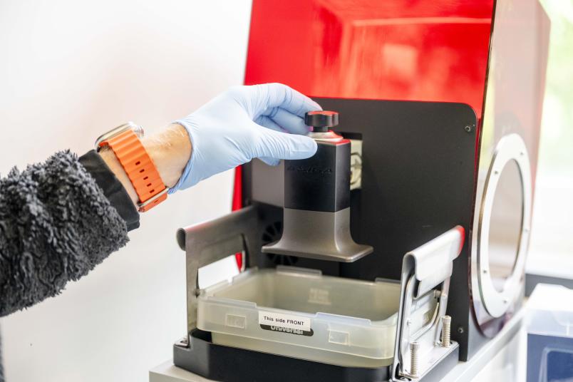
The 3D printing process has only recently become affordable for the production of suitable chips.
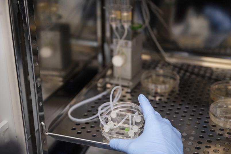
The cells that the researchers wish to observe are seeded onto the chips. The entire system is continuously flushed with nutrient solution via tubes.
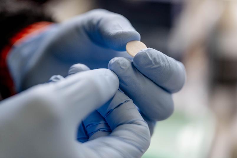
Such tiny chips, which are coated with bone substance, are seeded with human cells that break down and create bones.
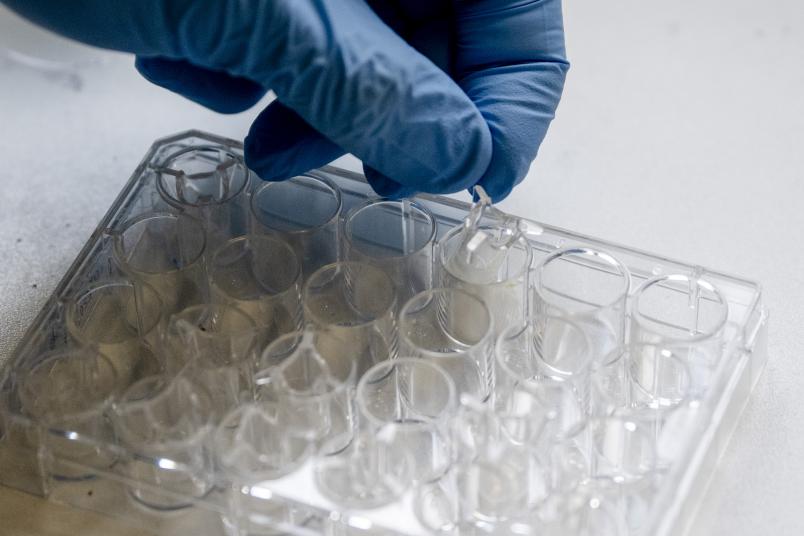
The cell culture can be observed to see how much bone substance is broken down or reformed.
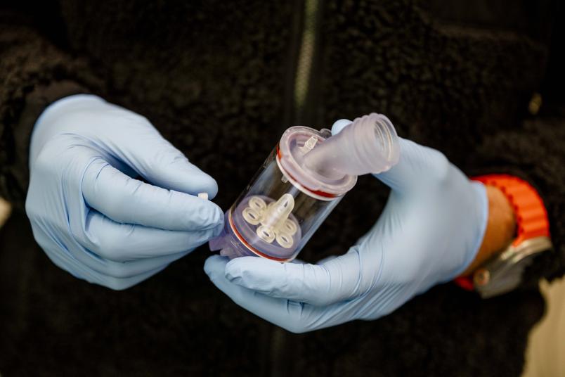
Larger samples can be placed in such bioreactors, where the researchers are able to observe cellular activity over several weeks.
There are multiple benefits to this approach. “We are avoiding unnecessary animal experimentation, and we receive data that are physiologically closer to humans than those from small animal models.” Differences in immune system, weight, strain, and joint mechanics make it difficult to translate results from small animal subjects. Furthermore, the cell culture system allows continuous microscopic observation and sensory analysis, which is either not possible in animal testing or places disproportionate strain on the animals. In this case, implantation and maintenance are performed, and examinations take place only at specific times.
3D-printed chips as an artificial bone environment
To make the cell cultures as realistic as possible, the researchers are developing their own scaffold structures. These are produced using 3D printers and biocompatible materials – a technology that has only recently become widely available. “A few years ago, this could only be financed for large industrial firms,” says Dr. Alexander Sieberath, a research assistant on the team. The printed chips are then coated with a fluorescent, bone-like substance. The team holds a patent for this procedure.
One dynamic system over a period of weeks
Once the chips are prepared, the researchers populate them with two central cell types: osteoblasts, which are obtained from precursor cells from bones or marrow, and osteoclasts, which are acquired from cells from donated blood. Osteoblasts build up bone substance, and osteoclasts break it down. The fluorescence of the chip coating makes it possible to track their activity in real time. Artificial intelligence aids in the quantitative assessment of the resorption processes.
The researchers also document the activity of the bone-forming osteoblasts, in particular the mineralization process, with an established histological coloration. This is visualized via a separate channel with a modern microscope and can be systematically documented for various points in time, resulting in a two-channel image of the bone metabolism: real-time resorption by osteoclasts and time-resolved mineralization by osteoblasts.
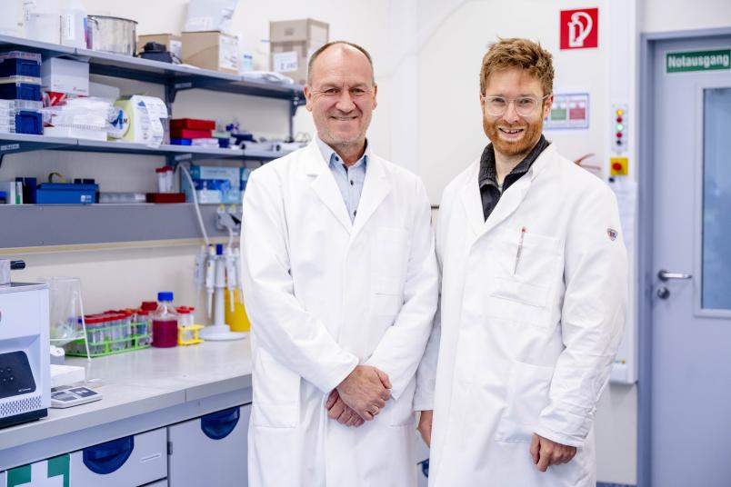
Jochen Salber (left) and Alexander Sieberath work with organoids instead of animal testing.
Tubes, pumps, and sensors create a microphysiological system that is continuously supplied with nutrient solution. “Such experiments can take place over several weeks,” explains Sieberath. “We can operate many chips parallel to one another and selectively adjust individual sets of parameters.” This allows, for example, for the determination of optimal drug dosages in high-throughput settings. “If it goes well, the formation of bones by osteoblasts comes out on top,” says Salber. “If the scales tip in favor of the osteoclasts, the bone becomes porous, which poses a risk to implant stability.”
An artificial organism
In one experiment, the researchers were already able to demonstrate the effect that the microstructuring of ceramic tooth implants has on bone formation and loss. They have also successfully used 3D printers to replicate bone structures – based on blueprints derived from CT scans of human bones – and to seed them with human cells. These larger models can also be studied in bioreactors that mimic conditions within the body. “It is even possible to replicate multiple organ systems using cells on chips and connect these organoids, creating a kind of artificial organism in which the organs influence one another,” says Salber. The chips, he adds, could also be equipped with sensors that constantly measure various parameters.
Projects and awards
Once we have explored the fundamentals of the interaction between bones and implant material, animal testing will still have to be conducted at some point to ensure safety before, say, an optimized implant is used in a clinical setting,” Salber adds. “We cannot entirely avoid this. But there is a difference between disposing of a cell culture when I make a mistake in the lab and sacrificing a test animal.”
Salber believes that the new approach methodologies to animal experimentation could be much improved. “The proper funding landscape isn’t in place at the moment,” he says. The further development of these highly realistic models requires, in particular, personnel resources—a point that poses a special challenge for a research group funded exclusively by third-party grants. Only with stable funding can these innovative, animal-free approaches be expanded and established in the long term.