Diagnosing cancer
New process for identifying biomarkers established
An early and precise cancer diagnosis is crucial for successful treatment.
Scientists at Ruhr-Universität Bochum have established a process for identifying biomarkers for the diagnosis of different types of cancer. With the aid of a specific type of infrared (IR) spectroscopy, the researchers applied an automated and label-free approach to detect tumour tissue in a biopsy or tissue sample. Unlike with label-based processes, such as are currently deployed by pathologists, the tissue remains unmodified. This, in turn, facilitates detailed protein analyses in the next step. Studying tissue samples from patients who suffered from lung or pleural cancer, the researchers identified protein biomarkers that are typical of the respective subtype of cancer.
The team of the research consortium “Protein Research Unit Ruhr within Europe” (PURE) has published their report in the journal “Scientific Reports”.
Specific workflow established
With their current work, the researchers from Bochum put their vision for PURE fully into practice. “For the first time, the workflow that has been planned from the outset has been completely reproduced,” explains Prof Dr Klaus Gerwert, Spokesman at PURE. In the process, the fresh tissue is instantly cooled, then brought to the study centre and documented. Afterwards, traditional diagnostics is carried out, which is standard for every patient, while simultaneously an analysis using the process made in Bochum is performed.
For the purpose of the study, the researchers worked with tissue samples of so-called diffuse malignant mesothelioma. They compared two mesothelioma subtypes – the sarcomatoid and epithelioid type. This type of cancer often scatters into the lung and is usually terminal.
Bochum-made process for diagnostics
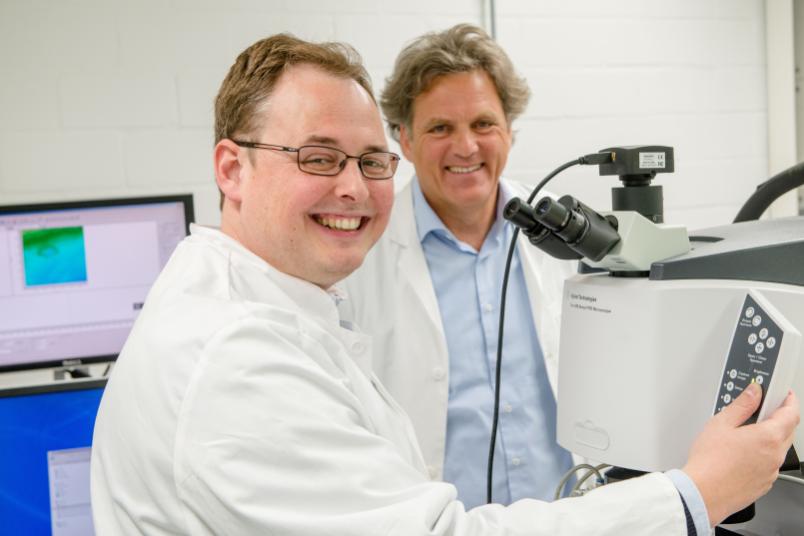
The researchers represented the spreading of a tumour with an IR microscope with high spatial resolution. Klaus Gerwert and Dr Frederik Großerüschkamp developed this imaging method under the umbrella of PURE. It enables them to distinguish between different subtypes of cancer – an important information for prognosis and therapy. It does not require any dye or antibodies, which are necessary in traditional pathology. Since the tissue remains unmodified, the same sample can subsequently be analysed on the molecular level.
After the tumour has been localised in the tissue sample, the researchers cut it out using special laser technology. At Medizinisches Proteom-Center at Ruhr-Universität, likewise a member of the PURE consortium, experts headed by Prof Dr Barbara Sitek and Prof Dr Katrin Marcus analyse the protein composition of the extracted tissue using mass spectrometry.
Personalised diagnosis
Thus, the researchers are able to determine which of the more than 2,000 identified proteins in the sarcomatoid mesothelioma are noticeably increased or reduced when compared to the epitheloid mesothelioma. “Consequently, we can determine changes in signal transduction in the cancer cells for each patient individually,” explains Klaus Gerwert. “This is crucial information for precise therapy.”
The thus identified proteins might in future be used as biomarkers, in order to detect the type of cancer in other patients. Researchers studied the correlation of the detected proteins and the biomarkers that are currently used in traditional pathology. The result: the method made in Bochum also identified the five biomarkers which are already used to diagnose the mesothelioma subtypes. “Thereby we have validated our method,” explains Gerwert. The PURE researchers also discovered additional biomarkers. Gerwert: “Those biomarkers are yet to be validated with a larger cohort in the next step.”
“At present, the newly developed label-free approach is unique, and it opens up the possibility to search specifically for biomarkers in future,” says Frederik Großerüschkamp.
The objective: finding biomarkers in body fluids
Gerwert and his colleagues intend to apply the method for other types of cancer and to thus identify new biomarkers. The Head of the Department of Biophysics outlines a prospect: “Our objective is to detect the biomarkers that we have now identified in tissue also in body fluids such as blood and urine,” says Gerwert. “This would facilitate non-invasive, precise and predictive diagnosis with the aid of a simple antibody test.”
Diffuse malignant mesothelioma
The diffuse malignant mesothelioma is primarily triggered due to exposure to asbestos. The tumours take very diverse forms, and a comparably small number of people are affected – this renders research quite difficult. “Especially in cases such as these, our new method might provide a valuable alternative in biomarker research,” elaborates Großerüschkamp.