Neuroscience
Fibromyalgia changes the brain
Pain-processing regions are particularly affected by changes in brain volume. The good news is that these changes may be reversible.
One of the core symptoms experienced by patients with fibromyalgia is chronic pain. A team from the LWL Clinic for Psychosomatic Medicine and Psychotherapy at Ruhr University Bochum, Germany, has investigated the brain changes that are related to the disorder. Using magnetic resonance data, the researchers proved that the areas of the brain involved in the processing and emotional evaluation of pain are altered in patients. This applies to the volume of both the grey matter, which mainly houses nerve cells, and the white matter, which mostly consists of fibre connections between the nerve cells. The researchers published their findings in the journal Arthritis Research and Therapy on 19 May 2023.
Changes in the pain network
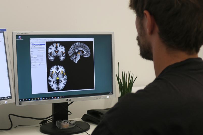
The team surrounding Professor Martin Diers and Benjamin Mosch analysed the magnetic resonance imaging data of 23 female patients with fibromyalgia and 21 healthy control subjects. They wanted to examine the volume of the grey matter, i.e. the nerve cells, in various pain-processing areas of the brain, and the so-called white matter, which mainly consists of the fibre connections between the nerve cells through which signals are transmitted. “One of our goals was to find out whether the directionality of the diffusion of water molecules differs in certain areas of the brain, in other words: whether we can identify any regional differences in signal transmission,“ explains Benjamin Mosch.
The researchers found changes of the grey matter volume mainly in the pain network of the brain, i.e. in the regions responsible for processing and evaluating pain. “In certain regions responsible for the inhibition of pain, we found a decrease in grey matter in the patients compared to the healthy individuals,” explains Benjamin Mosch. “In patients, the volume of these regions was significantly reduced.”
Regarding the transmission of signals, changes were found in the thalamus. The thalamus is considered as an important node in neuronal pain processing. The deviations of the white matter in patients with fibromyalgia compared to healthy controls indicate an altered conduction of pain signals in patients with fibromyalgia.
Relationships between brain structure, perception and behaviour
The team finally related the results of the structural brain changes to perceptional and behavioural characteristics of the study participants. The amount of decreased volume in a number of relevant brain regions is inversely related with the amount of perceived pain the patients report. The researchers made an interesting observation when analysing the correlation between depressiveness or activity levels with the change in the volume of certain brain areas. The volume of the so-called putamen correlated negatively with the expression of depressive symptoms and positively with the activity level of the participants. “This indicates that changes in the brain may not be permanent, but that they can be influenced; in other words they might be reversible, for example through an active everyday life,” concludes Benjamin Mosch.