
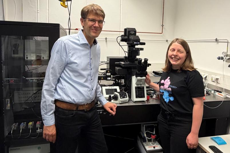
Simon Ebbinghaus and Mailin Becker (right) have developed a new imaging technique.
Imaging
Researchers Compare Protein Folding within Cells
A new method makes it possible to measure the stability of proteins at various points within an individual cell. This sheds more light on the development of neurodegenerative diseases.
Protein aggregates are associated with the development of diseases like Alzheimer’s and Parkinson’s. These are found more often in certain areas of cells known as membraneless organelles. Do these organelles promote this protein clumping, or do they protect the cell by temporarily absorbing the aggregates? Professor Simon Ebbinghaus and Mailin Becker from Ruhr University Bochum, Germany, set out to answer this question with their new, self-developed method “confocal Fast Relaxation Imaging,” or cFReI. They report their findings in the journal PRX Life from July 16, 2026.
Proteins and RNA accumulate
Proteins are involved in numerous processes within the cell. In order for all of these to flow smoothly, the cell is divided into different compartments known as organelles. Some of these, such as the nucleus or the mitochondria, are surrounded by a membrane. “However, there are also organelles that are not encased by a membrane,” says Becker. Proteins and RNA accumulate in these membraneless organelles (MLOs). “They are formed through liquid-liquid phase separation,” she explains. “This process is similar to the formation of oil droplets in water.”
Many of these MLOs contain proteins that fold incorrectly, or unravel and then clump up. The resulting protein aggregates are linked to neurodegenerative diseases like amyotrophic lateral sclerosis (ALS), Alzheimer’s, or Parkinson’s. However, it is not yet clear what role MLOs play in the development of these diseases. On the one hand, MLOs could protect the cell by temporarily absorbing misfolded proteins; yet they could also be places that promote the misfolding of proteins and thereby harm the cell. “In order to understand how these diseases develop, it is important to investigate the unfolding of proteins directly within the complex environment of living cells,” says Ebbinghaus.
Direct comparison of the stability of proteins within a cell
For this purpose, the researchers at Ruhr University Bochum and the Research Center Chemical Sciences and Sustainability have developed a novel experimental method known as “confocal Fast Relaxation Imaging,” or cFReI. This makes it possible to directly compare the stability – and, thus, the unfolding – of proteins within MLOs and in the surrounding cytoplasm of the same cell.
Using this method, they examined a variant of the protein superoxide dismutase (SOD1) that accumulates in stress granules. Stress granules are MLOs that form in the cytoplasm as a reaction to cellular stress and help the cell overcome it.
Observation provides surprising insight
“Contrary to our expectations from in-vitro experiments, we observed that SOD1 is not destabilized within the stress granules,” reports Becker. “In fact, the protein actually stabilized within the stress granules in most cells.” This, she says, indicates that MLOs could protect the cell against misfolded proteins by temporarily absorbing them instead of promoting their misfolding.
The new method is not restricted to SOD1 or stress granules – it can also be applied to other biomolecules in different organelles, thereby providing important insight into the development of neurodegenerative diseases. In the long term, it could also reveal new starting points for therapeutic strategies.