
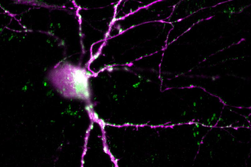
The purple structure shows a nerve cell that has been stained with the dye “mCherry”. The GIRK channels are visible in green.
Neurobiology
New Optogenetic Tool Inhibits Neurons and Heart Muscle Cells
Researchers hope the tool will help advance research into heart rhythm disorders and other diseases.
Researchers from Ruhr University Bochum have partnered with colleagues in Göttingen, Berlin, Paris, and Essen to develop new optogenetic tools that can selectively inhibit cellular activity through light. These tools could help investigate diseases in which the balance between excitation and inhibition is disrupted, such as cardiac arrhythmias. The team working alongside Dr. Brix Mücher, Dr. Ida Siveke, and Professor Stefan Herlitze from the Department of General Zoology and Neurobiology at Ruhr University Bochum, Germany, reports its findings in the journal Science Signaling, published online on October 20, 2026.
Optogenetic tools can be used to activate and deactivate nerve cells via light. In their present study, the researchers coupled a light-sensitive receptor named Opn4L to a modified potassium channel named GIRK1, which activates a G-protein-coupled signaling pathway.
G-protein-coupled receptors are among the most important signal receptors in animal cells. They respond to various stimuli, including signaling molecules, medications, and light. In addition, they can activate different G-proteins depending on cell and receptor type, thereby triggering various responses. Serotonin and dopamine receptors are typical examples: Disruptions to these signaling pathways play a key role in depression, schizophrenia, and Parkinson’s disease.
Test with neurons and muscle cells
In cell culture experiments, the researchers introduced the newly developed optogenetic tool, based on Opn4L/GIRK1, into cerebellar neurons and heart muscle cells. The experiments with cerebellar cells demonstrated that when blue light shines onto the Opn4L receptor, the coupled GIRK channel in the cell membrane opens, activating a downstream G-protein-coupled signaling pathway. This makes the cell less excitable; therefore, the activity of the neurons is inhibited by the activation of the optogenetic tool.
This optical switch also functioned in human heart muscle cells. When the researchers exposed the Opn4L/GIRK1 construct to blue light, the spontaneous beating rate of the cultured cells decreased.
“We have developed an optogenetic tool that not only activates a receptor, but also selectively reinforces its downstream signaling pathway,” explains Ida Siveke from Ruhr University Bochum. “This allows us to very precisely influence the balance between stimulating and inhibiting signals in individual cells.”
“As a result, the new optogenetic constructs could help us more accurately examine and control the underlying signaling mechanisms in heart muscle and pacemaker cells as well as neurons in the future,” says optogenetics pioneer Professor Stefan Herlitze. “This opens up entirely new areas of application for optogenetics.”