Neuroscience
When the cerebellum becomes active
A new study shows that the cerebellum is involved in processing emotions. This is important to know when caring for people with ataxia.
For a long time, the fact that the cerebellum also plays an important role in regulating our emotions – such as when processing fear – has been ignored. Professor Melanie Mark from Ruhr-University Bochum and Professor Dagmar Timmann from the University of Duisburg-Essen are two of the first researchers to provide experimental evidence that the cerebellum contributes towards both the learning and the extinction of conditioned fear responses. They report on this in the journal eNeuro on January 4, 2024.
To investigate the role that the cerebellum plays in fear learning, the two neuroscientists conducted learning experiments – the neurologist with humans and the neurobiologist with mice. “In our studies, we draw on classic fear-conditioning experiments and compare healthy humans and mice with those that have a cerebellar disease, ataxia,” says Timmann, summarizing the shared study design.
To understand ataxia
In her latest study, the clinical neuroscientist selected 20 subjects who suffer from rare cerebellar diseases such as spinocerebellar ataxia type 6 (SCA6), in addition to 20 healthy people. “The movement disorder SCA6 is triggered by a genetic defect similar to Huntington’s disease and only affects a very small number of people in Germany,” explains Timmann, who has been offering ataxia consultations at Essen University Hospital for many years. “SCA6 is associated with a loss of a specific type of neuron in the cerebellum, the Purkinje cells. The Purkinje cells are important as intermediaries between the cerebellum and the rest of the brain. For instance, the cerebellum helps the cerebrum to optimize motion sequences,” says the researcher.
Fear-conditioning experiments
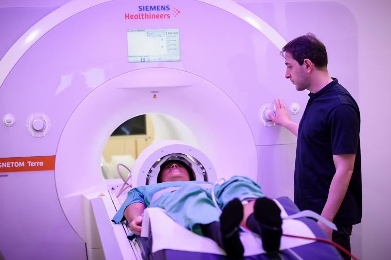
In their study, Timmann and her team had patients and participants learn and then unlearn fear within two days while observing them in the 7 Tesla MRI scanner.
The direct comparison of healthy subjects and people with ataxia confirmed the assumption that people with ataxia have deficits when learning and unlearning fear. Not only did the acquisition and consolidation of the learned fear response take longer than in the healthy control group, but unlearning the fear was also more prolonged. However, the deficits were much lower than expected: “Beforehand, we assumed that our ataxia patients would be much more significantly impaired during the fear conditioning and that this, in turn, would be associated with clearly visible changes in the cerebellum,” says Timmann. However, in the 7 Tesla MRI, the activation pattern in the ataxia patients was also reasonably well preserved and only showed minor deviations from the healthy participants.
Mouse models
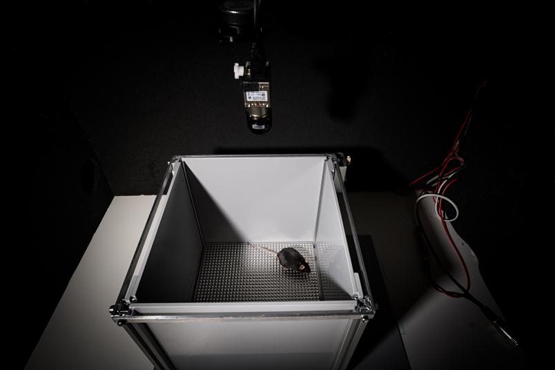
To confirm the Essen clinician’s observations, her research colleague in Bochum, neurobiologist Melanie Mark, conducted the fear conditioning study with healthy mice and mice suffering from SCA6. Mark used SCA6 mouse models that she had already developed for previous studies.
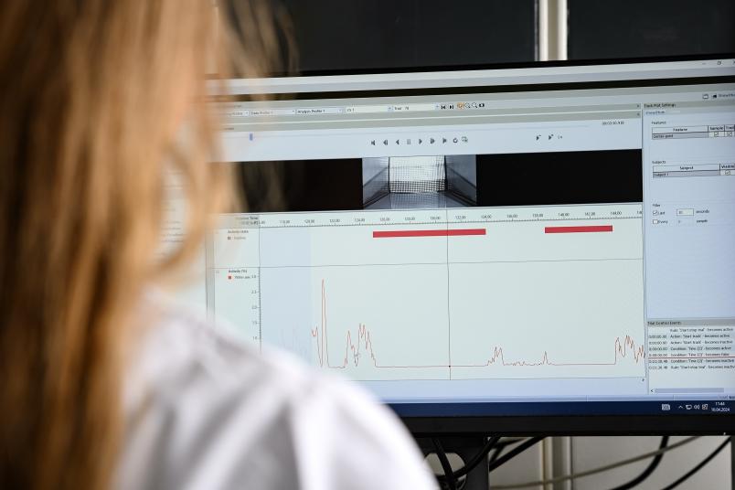
In the experiment, the healthy and sick mice first learned to associate a tone with an unpleasant electric shock and then unlearned the association. “Our SCA6 mice were able to learn the fear response exactly like the people with ataxia, but they did not consolidate what they had learned. Their memory of the learned association task did not last until the next day,” explains Mark. The researcher was thus able to show that the fear memory in the SCA6 mice was disrupted in comparison to the healthy mice. The cerebellar disease prevented the mice from consolidating what they had learned and, based on this, from being able to make a learned prediction.
Mark thus came to the same conclusion as Timmann: The cerebellum plays a role in learning fear responses. However, the deficits were also much lower than expected in the mouse model. “In this chronic illness, other regions of the brain have possibly learned to compensate for the cerebellar deficit. This is desirable from an evolutionary perspective. If a region fails, the whole neuronal circuit does not immediately collapse. This does not mean that the cerebellum is not involved,” explain Mark and Timmann. Melanie Mark’s team is now working hard on rectifying the learning deficits in SCA 6 mice using various methods.


